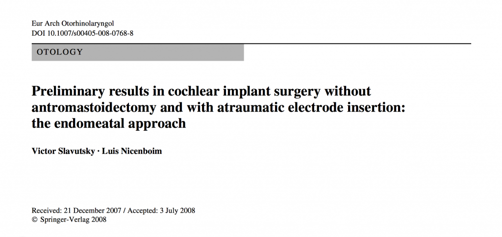
El abordaje endomeatal para el implante coclear.
EMA es una via de elección en esta cirugía. El Implante coclear se realiza generalmente en oídos sanos. La patología afecta solamente la cóclea y no compromete el resto del hueso temporal.
EMA siempre accede a la ventana redonda (VR), que no es posible por timpanotomía posterior, en mas de un 30% de los casos. La VR es un orificio natural que hace innecesaria la cocleostomía promontorial, evita la falsa víxa y garantiza una inserción atraumática del haz de electrodos en la escala timpánica (ET).
EMA conserva el MACS y su función, que mantiene la presión del oído medio y facilita la transmisión acústica en los Implante Hibridos, favoreciendo el resultado funcional.
EMA no pone en riesgo el nervio facial, porque no es una referencia anatómica en este abordaje.
EMA es una técnica mínimamente invasiva que permite una cirugía en plan ambulatorio. La destrucción de la mastoides no solo restringe el acceso a la VR, sino que tampoco permite una linea de inserción que coincida con la proyección del eje de la escala timpánica. La timpanotomía posterior es una ventana anatómica que no solo precisa de exponer el nervio facial, sino que es un espacio quirúrgico estrecho que condiciona y determina el sitio de la cocleostomía promontorial y es el motivo por el cual no existe consenso sobre el lugar en que debe practicarse.
EMA utiliza como via de acceso a la cóclea el meato auditivo externo, un espacio anatómico mas amplio, que permite orientar con libertad la guía de electrodos hacia la VR. De un orificio natural a otro orificio natural, con todas las ventajas que comporta.
No tiene sentido quirúrgico la destrucción de tejidos sanos sino se alcanza un mejor resultado funcional.
ENDOMEATAL APPROACH FOR COCHLEAR IMPLANT
EMA is an elected technique for this surgery. The cochlear implant is usually performed in healthy ears. The disease affects only the cochlea and does not compromise the rest of the temporal bone.
EMA always accesses the round window (RW), which is not possible for posterior tympanotomy , in over 30% of cases. The RW is a natural orifice and promontorial cochleostomy becomes unnecessary , prevents false pathway and ensures atraumatic insertion of the electrode bundle in the scala tympani (ST) .
EMA preserves the MACS and its function, which keeps the middle ear pressure and facilitates the acoustic transmission in Hybrid Implant , favoring functional outcome.
EMA does not take facial nerve risk, because it is not an anatomical reference in this approach.
EMA is a minimally invasive technique that allows outpatient surgery. Transcortical mastoid approach not only restricts access to the RW membrane , also the insertion line of the electrodode lead does not matches the projection of the axis of the scala tympani . The posterior tympanotomy is not only accurate to expose the facial nerve it is also a narrow surgical space that determines the site of the promontorial cochleostomy and is the reason why there is no consensus on where it should be practiced.
EMA approach the cochlea through the external auditory meatus , a wider anatomical space , which allows direct the electrode lead to the RW . From a natural orifice to another natural orifice, with all the advantages that entails.
To destroy healthy tissues does not have any surgical sense If a better functional outcame is not reached.
Tesis Doctoral. Universidad Autónoma Barcelona – Victor Slavutsky (clik)
ATLAS DE CIRUGIA OTOLOGICA. Capítulo de Implantes Cocleares.(Pgs.16-38)
 Endomeatal approach for Cochlear Implant
Cochlear Implant surgery through natural orifices. The endomeatal approach
EMA – Francesco Galletti et al.
Endomeatal approach in cochlear implant surgery in a patient with small mastoid cavity and procident lateral sinus
Università degli Studi di Messina
Francesco Galletti, Francesco Freni, Francesco Gazia, Bruno Galletti
Images in…
To cite: Galletti F, Freni F,Gazia F, et al. BMJ CaseRep 2019;12:e229518.
doi:10.1136/bcr-2019-229518
Department of Adult and
Development Age Human Pathology “Gaetano Barresi”,
Unit of Otorhinolaryngology,Universita degli Studi di Messina, Messina, Italy
Correspondence to
Dr Francesco Gazia,
ssgazia@ gmail. com
Accepted 21 May 2019
© BMJ Publishing Group
Limited 2019. No commercial re-use. See rights and permissions. Published by BMJ.
Description
Endomeatal approach (EMA) in cochlear implant
(CI) surgery avoid mastoidectomy and posterior
tympanotomy (PT) by using the external auditory
canal (EAC) and the round window (RW) as
a natural access pathway for CI positioning.1 We
present a modified technique for EMA applied in a
78-year-old patient with profound hearing loss and
a history of diabetes and hypothyroidism.2 3 For
several years, the patient has reported poor improvements
through the use of an auricular prosthetic
device. Our multidisciplinary team decided to
undertake the procedure and perform a CI surgery
in the ear with a worse auditory performance. The
patient underwent vaccination for Streptococcus
pneumoniae, Haemophilus influenzae and Neisseria
meningitidis, and CT of the brain and MRI
have been performed. The instrumental diagnosis
displayed the presence of a small mastoid cavity and
a bilateral procident lateral sinus.4
Given the radiological objective, it was decided
to perform CI surgery with an EMA, whose surgical
steps are detailed on video (Video 1).
A month after the surgery, CI was correctly activated,
with no signs of postsurgical complications
being displayed. The patient showed an excellent
capacity for social interaction and a great fitting
of the implant.
The incidence of facial nerve lesions following
PT—which are mainly caused by direct injury or
heat produced by drilling—has been reported to
be 1%.5 The visualisation of the facial channel over
the platina of the stape provided a safely controlled
technique to ensure a healthy formation of the
groove and avoid the occurrence of alterations to
the facial nerve—located in the posterior EAC wall.
Thanks to mobilisation of the incus, we are sure to
maintain the facial nerve intact.
The main advantages of this technique compared
with the traditional mastoidectomy and PT are
recognised as follows:
A faster better access to the scala tympani.
A wider electrode insertion angle.
A decreased risk for the groove formation to
damage the facial nerve, as it is placed in the
posterior EAC wall, which is distant to the
nerve.
Avoidance of cholesteatoma and middle ear
infections since only a small segment of the electrode
array resides in the middle ear space, and
no mastoid cavity is present.
A drastic reduction in the risk for the electrode
to get in contact with the skin and the extrusion,
as the bone paté is enough to support the electrode
inside the groove.
EMA discourages false pathways and increases
stimulation of the neuronal population by
placing the basal electrodes at the onset of the
scala tympani.
EMA is a soft surgery technique that facilitates
CI collocation in alterations of classical anatomy,
such as anteriorly located facial nerve, procident
lateral sinus, narrow facial recess and small
mastoid cavity.
Given the excellent result of our case and the
possibility for this technique to be used on craniofacial
malformations, we deem EMA to be an
effective, safe and minimally invasive technique
even in anatomically varying cases affecting the
ear.
Learning points
►► Endomeatal approach (EMA) in cochlear
implant surgery should be the gold standard in
anatomically varying cases, such as procident
lateral sinus and small mastoid cavity.
►► The changes made to the traditional EMA allow
a better preservation of the facial nerve and a
lower occurrence of dislocation of the array.
Contributors FGal was the first surgeon. BG and FF were the
assistants. FGaz made the video and prepared the manuscript.
Funding The authors have not declared a specific grant for this
research from any funding agency in the public, commercial or
not-for-profit sectors.
Competing interests None declared.
Video 1 Surgery steps in endomeatal approach in
cochlear implant collocation
BMJ Case Rep: first published as 10.1136/bcr-2019-229518 on 6 June 2019. Downloaded from http://casereports.bmj.com/ on 6 June 2019 by DrFrancesco Gazia. Protected by copyright.
2 Galletti F, et al. BMJ Case Rep 2019;12:e229518. doi:10.1136/bcr-2019-229518
Images in…
Patient consent for publication Obtained.
Provenance and peer review Not commissioned; externally peer reviewed.
References
1 Slavutsky V, Nicenboim L. Preliminary results in cochlear implant surgery without antromastoidectomy and with atraumatic electrode insertion: the
endomeatal approach. Eur Arch Otorhinolaryngol
2009;266:481–8.
2 Freni F, Galletti B, Galletti F, et al. Improved outcomes for papillary thyroid
microcarcinoma care: active surveillance and case volume. Ther Adv Endocrinol Metab
2018;9:185–6.
3 Galletti B, Bruno R, Catalano N, et al. Follicular carcinoma on a radio-treated ectopic
lingual thyroid. Chirurgia 2016;29:88–91.
4 Ciodaro F, Freni F, Mannella VK, et al. Use of 3D Volume Rendering Based on High-
Resolution Computed Tomography Temporal Bone in Patients with Cochlear Implants.
Am J Case Rep 2019;20:184–8.
5 El-Anwar MW, ElAassar AS, Foad YA. Non-mastoidectomy Cochlear Implant
Approaches: A Literature Review. Int Arch Otorhinolaryngol 2016;20:180–4.
Copyright 2019 BMJ Publishing Group. All rights reserved. For permission to reuse any of this content visit
BMJ Case Report Fellows may re-use this article for personal use and teaching without any further permission.
+44 (0) 207111 1105 or via email at support@bmj.com.
Visit casereports.bmj.com for more articles like this and to become a Fellow
BMJ Case Rep: first published as 10.1136/bcr-2019-229518 on 6 June 2019. Downloaded from http://casereports.bmj.com/ on 6 June 2019 by DrFrancesco Gazia. Protected by copyright.
View publication stats

